CT and Magnetic Resonance Imaging
The CT and MRI Department first became operational in 1992, with the installation of the first CT scanner in a Pediatric Hospital. In 1999, the first MRI scanner was installed, followed by a second one in 2013.
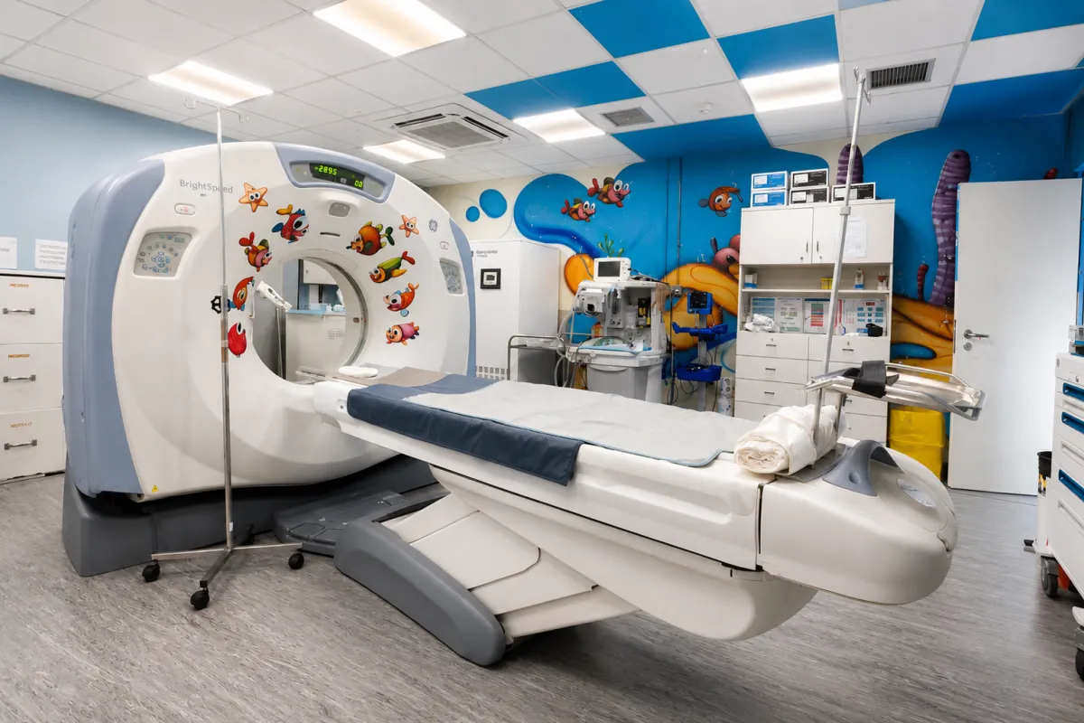
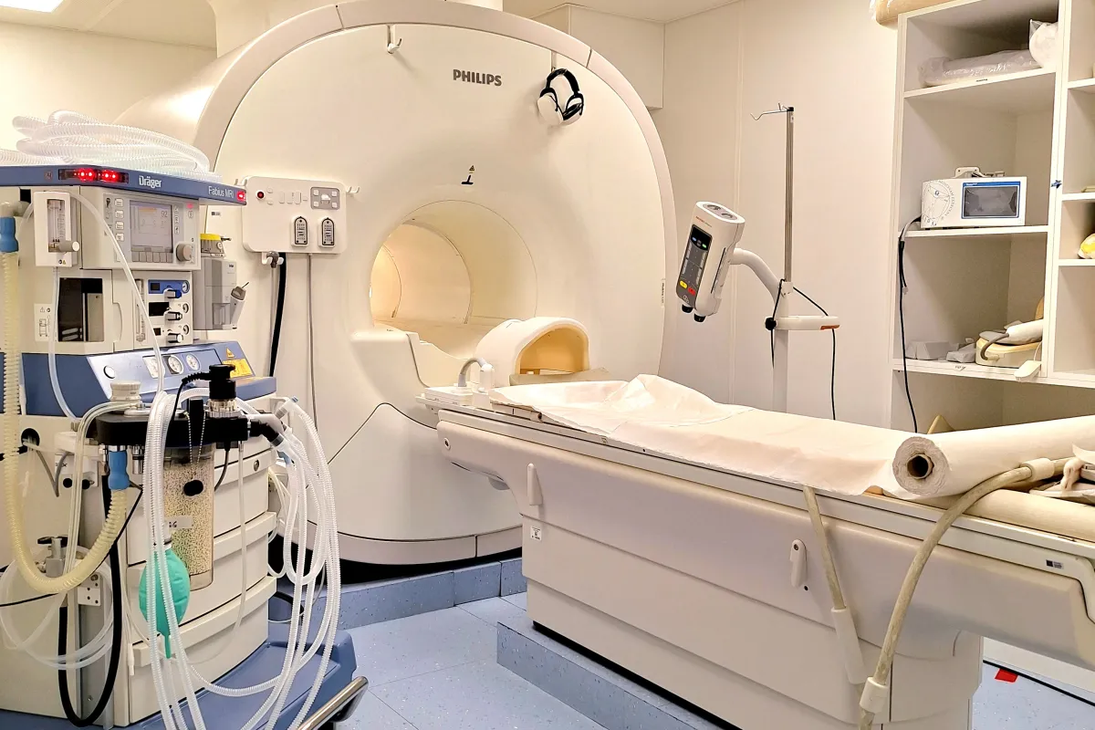
Today, the Department is equipped with 2 state-of-the-art 1.5 Tesla MRI scanners featuring artificial intelligence applications, as well as a multislice CT scanner.
Today, the Department is equipped with 2 state-of-the-art 1.5 Tesla MRI scanners featuring artificial intelligence applications, as well as a multislice CT scanner.
Coverage.
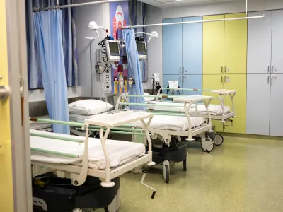
Provides coverage for approximately 900 hospital beds at the “Agia Sofia” and “P. & A. Kyriakou” Children’s Hospitals, oncology patients at “ELPIDA”, as well as outpatient cases involving newborns, infants, and children referred from other hospitals (Penteli Children’s Hospital, Attikon, Alexandra, Nikaia, Elena, and regional hospitals).
Εικόνα
Εικόνα
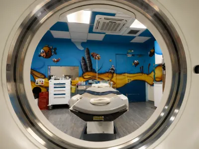
Reduced radiation dose protocols are applied in pediatric patients, following the European standard for diagnostic reference levels (PDRL) based on RP No. 185.
Εικόνα
Typical Examinations
- Brain
- Orbits
- Petrous bones
- Paranasal sinuses
- Facial skeleton
- Neck
- Chest
- Cervical spine
- Thoracic spine
- Lumbar and sacrococcygeal spine
- Upper abdomen
- Retroperitoneal space
- Lower abdomen (pelvis)
- Bones and soft tissues
Special Examinations & Interventional Procedures
- CT angiography of the brain, neck, chest, abdomen and all body regions
- CT urography / pyelography
- Tissue, soft tissue and bone biopsies
- Treatment of bone lesions (ablation or thermocoagulation / cryoablation)
- Drainage of collections
Special Examinations & Interventional Procedures
- CT angiography of the brain, neck, chest, abdomen and all body regions
- CT urography / pyelography
- Tissue, soft tissue and bone biopsies
- Treatment of bone lesions (ablation or thermocoagulation / cryoablation)
- Drainage of collections
Use of artificial intelligence and deep learning algorithms (AirRecon DL) in image reconstruction — reducing examination time by up to 50% while simultaneously improving image quality. Use of new soft “blanket-type” AIR COILS with full adaptation to each child’s anatomy, as well as an integrated video, audio and music system.
Εικόνα
Typical Examinations
- Brain
- Pituitary gland
- Orbits
- Petrous bones
- Paranasal sinuses
- Facial skeleton
- Neck
- Chest
- Cervical / thoracic / lumbar & sacrococcygeal spine
- Upper abdomen
- Retroperitoneal space
- Lower abdomen (pelvis)
- Internal genital organs
- Magnetic Resonance Cholangiopancreatography (MRCP)
- Bones, soft tissues and joints
- MRI Neuronavigation (MRI NAVI)
Special Examinations
- Brain diffusion and perfusion MRI
- Brain MR spectroscopy
- Brain MR tractography
- Functional brain MRI
- MR enterography
- MR urography
- Measurement of iron deposition in the liver
- MR angiography (brain, neck, chest, abdomen)
- Whole-body MRI
Before the examination, children can play with an MRI simulator and become familiar with the procedure.
Examinations Under Anesthesia
In collaboration with the Hospital’s anesthesiologists, diagnostic examinations and invasive procedures are regularly performed on young children (inpatients & outpatients) who require sedation.
Preparation Instructions
- The child must not have had a cold, fever, cough, runny nose, vomiting, or diarrhea during the previous week or on the day of the examination.
- At least 40 days must have passed since an infectious illness (chickenpox, measles, rubella, etc.) and 20 days since any vaccination.
- The night before: a light meal before 21:00, milk until midnight, and water no later than 5 hours before the examination. For newborns/infants up to 1 year of age: breastfeeding until 03:00 and formula milk until 02:00.
- A recent complete blood count (CBC), performed within the last 7 days, must be provided.